
Melasma vs Lentigo vs PIH vs ABNOM: How to Tell Them Apart
Brown facial marks look alike but are different diseases, and a wrong diagnosis makes them worse. A Seoul dermatologist's guide to distinguishing melasma, solar lentigo, PIH and Hori's nevus.
This is an English adaptation of a clinical article Dr. SangYoul Yun (board-certified dermatologist and Medical Director of Delight Dermatology in Gangnam, Seoul), originally published in Korean. Read the Korean original on Naver. It has been restructured and translated for international readers; all references are the author's own.
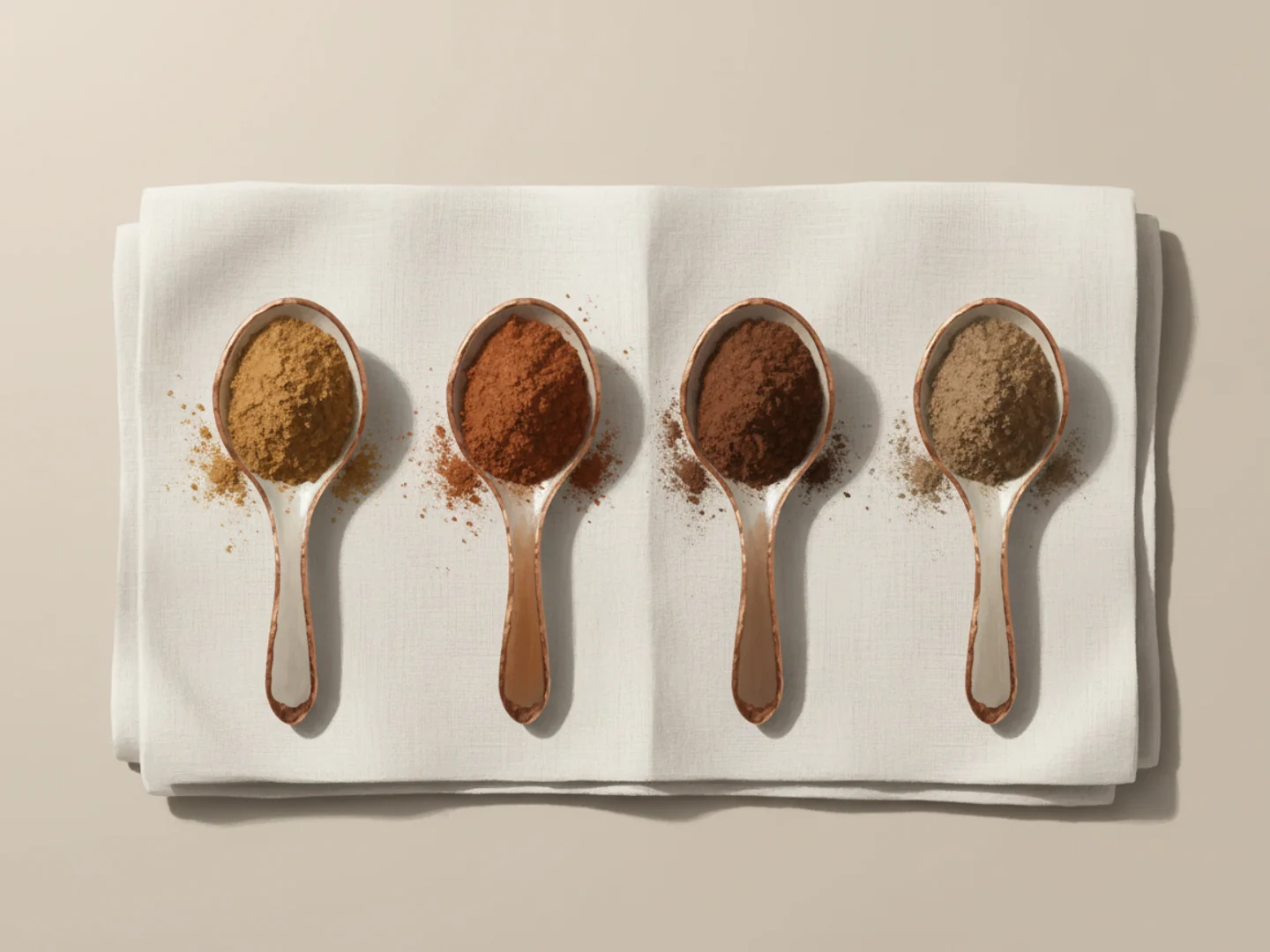
"I read online and got a melasma laser, but they say it didn't work, that it wasn't actually melasma?" I hear this often in clinic. Yes, exactly. Brown marks on the face look similar on the surface but are completely different medical conditions, and a wrong diagnosis means treatment either fails or makes things worse.
Melasma, solar lentigo, post-inflammatory hyperpigmentation (PIH), and ABNOM (acquired bilateral nevus of Ota-like macules) are the four most common (and most often confused) pigment disorders in Korean women. Here is how to distinguish them, with the clinical diagnostic criteria.
1. Why accurate differentiation matters
The bottom line first: effective treatments differ completely depending on the disorder. Melasma needs gentle toning with adjunct medication, and using lentigo treatments (strong IPL or high-power lasers) makes melasma worse.1 ABNOM, with pigment in the dermis, barely responds to superficial lentigo treatments,2 and PIH recurs after a procedure unless the causative inflammation is controlled first. So the first thing we do in clinic is diagnose exactly which pigment disorder this is: if the diagnosis is wrong, treatment inevitably fails.
2. The four main pigment disorders, distinguished
① Melasma
- Location: symmetric on the cheeks, forehead, upper lip; spares the eyelids3
- Shape: brown patches with blurred borders (irregular, large areas)
- Cause: multifactorial (hormones, UV, heat, pregnancy, oral contraceptives, chronic inflammation)on
- Dermoscopy: uniform yellow-brown pigment plus a fine capillary network (seen in about 74%)4
- Wood's lamp: epidermal type darkens; dermal type shows little change4
- Key clue: symmetry and a link to hormonal change (onset/worsening after pregnancy or the pill)
② Solar lentigo
- Location: asymmetrically scattered on sun-exposed areas (forehead, temples, cheeks, backs of hands)
- Shape: small brown macules with sharp borders (usually under 5 mm)
- Cause: chronic UV exposure (photoaging) is the biggest driver
- Wood's lamp: darkens more distinctly (because it is epidermal pigment)4
- Key clue: once formed, it doesn't fade without sun protection (persists even in winter)5
Note: freckles (ephelides) are also small brown macules, but they darken in summer and fade in winter, distinguishing them from solar lentigo. Freckles appear in childhood and have a strong genetic component.5
③ Post-inflammatory hyperpigmentation (PIH)
- Location: exactly where prior inflammation, trauma, or acne occurred
- Shape: brown to gray-brown deposition in the shape of the former inflammation
- Cause: the aftermath of any skin inflammation (acne, eczema, dermatitis, peels, laser procedures)
- Dermoscopy: uniform brown or gray-brown pigment1
- Key clue: a distribution matching the inflammatory lesions (acne-scar sites, mosquito bites, etc.)
Caution: the darker the skin (Fitzpatrick IV–VI), the far higher the rate of PIH.1 Korean skin is prone to PIH, so minimizing inflammation is central to any procedure.
④ ABNOM (acquired nevus of Ota-like macules / Hori's nevus)
- Location: symmetric on both cheekbones, temples, and beside the nose; does not involve mucosa (eyes, mouth)6
- Shape: clustered blue-gray to blue-brown dots
- Cause: melanocytes located in the deep (mid) dermis (acquired dermal melanocytosis)6,7
- Wood's lamp: unlike superficial pigment, it does not darken further under Wood's lamp (deep dermal pigment)
- Key clue: mainly found in women in their 30s–40s6, a bluish hue (melasma is yellow-brown), and (being deep in the dermis) barely responds to superficial treatments like toning
Clinical tip: ABNOM is the most common acquired dermal pigment disorder in Korean, Japanese, and Chinese women. Very often patients come in after being treated for "melasma" with no result.
3. At-a-glance differential table
| Feature | Melasma | Lentigo | PIH | ABNOM |
|---|---|---|---|---|
| Distribution | Symmetric patches | Scattered on sun-exposed areas | Sites of inflammation | Symmetric on cheekbones/temples |
| Shape | Large area, blurred border | Small dots, sharp border | Shape of the inflammation | Small blue dots |
| Color | Yellow-brown | Brown to dark brown | Brown to gray-brown | Blue-gray / blue-brown |
| Depth | Epidermal–dermal mixed | Epidermal | Epidermal–dermal | Deep dermis |
| Wood's lamp | Epidermal type darkens | Darkens | Superficial darkens | Little change |
| Main cause | Hormones, UV | Chronic UV | Prior inflammation | Acquired dermal melanocytes |
| Typical age | Women 20s–40s | Middle-aged to older | All ages | Women 15s–40s |
4. How we diagnose: the clinic's pigment tools
For pigment-disorder care we use the following tools together.
- Wood's lamp: uses a UV wavelength to assess pigment depth. Epidermal pigment appears darker; dermal pigment shows little change or looks fainter.8
- Dermoscopy: 10× magnification is used to observe pigment patterns directly, distinguishing melasma's capillary network, lentigo's sharp border, and ABNOM's gray-blue dots.4
- META VIEW skin analysis: multi-wavelength imaging quantifies surface pigment, deep pigment, erythema, and UV damage.
- Clinical history: a full review of onset, hormonal change, pregnancy, medication use, and prior inflammation.
5. Different diagnosis, completely different treatment
| Disorder | Recommended treatment direction |
|---|---|
| Melasma | Low-irritation toning + adjunct drugs such as tranexamic acid + strict sun protection (strong lasers contraindicated)1 |
| Lentigo | Superficial pigment-targeting procedures such as pico / QS laser + sun protection |
| PIH | Treat the causative inflammation first + anti-inflammatory topicals + low-irritation procedures |
| ABNOM | Dermal-pigment-targeting lasers such as 1064 nm Q-switched Nd:YAG (multiple sessions needed)2,6 |
Even for marks of the same brown color, the treatments are often opposite. Melasma and lentigo look alike in color but require opposite treatment approaches, and using strong IPL intended for lentigo on melasma darkens the melasma.1
6. The most common misdiagnoses
- ABNOM mistaken for melasma: bluish pigment on both cheekbones diagnosed as "melasma" and toned repeatedly with no improvement. It is actually ABNOM, dermal pigment that shallow toning can't reach.
- PIH mistaken for melasma: brown acne marks lingering and mistaken for melasma. It is actually PIH; the acne inflammation must be controlled first, with anti-inflammatory treatment taking priority.
- Dermal melasma mistaken for lentigo: a small, relatively sharp-bordered melasma patch treated as lentigo with strong IPL → melasma worsens with accompanying PIH.
- Melasma–ABNOM mixed: one patient often has both, requiring the appropriate treatment for each in stages.6
7. How we approach pigment at Delight Dermatology
Diagnosis first, treatment second: a full assessment with dermoscopy and META VIEW. Because one patient can have several disorders together, we account for mixed types, and because Korean skin is at high risk of PIH, we prioritize low-irritation approaches. Since most pigment disorders recur, we approach treatment as long-term "management" rather than a "cure."
Bottom line: not all "brown marks" are the same. Melasma, lentigo, PIH, and ABNOM look alike but are separate diseases with completely different causes, depths, and treatments. Before choosing a procedure based on online information or advertising, get an accurate differential diagnosis.
References
- Honigman A, Lim AC, Murase JE. Differential diagnosis of melasma and hyperpigmentation. Dermatological Reviews. 2023.
- Ee HL, Goh CL, Khoo LS, et al. Treatment of acquired bilateral nevus of Ota-like macules (Hori's nevus) with a combination of the 532 nm Q-switched Nd:YAG laser followed by the 1064 nm Q-switched Nd:YAG is more effective: prospective study. Dermatol Surg. 2006;32(1):34-40.
- Passeron T, Picardo M. Melasma, a photoaging disorder. Pigment Cell Melanoma Res. 2018;31(4):461-465.
- Wang L, Xu AE. Dermoscopy combined with Wood lamp, a diagnostic alternative for five pigmented lesions on the face: an observational study. Chin Med J (Engl). 2020.
- Praetorius C, et al. Ephelides and lentigines: clinical and genetic features. DermNet NZ. 2023.
- Hori Y, Kawashima M, Oohara K, Kukita A. Acquired, bilateral nevus of Ota-like macules. J Am Acad Dermatol. 1984;10(6):961-964.
- Cho SB, et al. Treatment of acquired bilateral nevus of Ota-like macules (Hori's nevus) using 1064-nm Q-switched Nd:YAG laser with low fluence. Int J Dermatol. 2009;48(12):1308-1312.
- Sonthalia S, et al. Evaluation of Dermoscopic Features in Facial Melanosis with Wood Lamp Examination. Clin Cosmet Investig Dermatol. 2022;15:215-227.
Medical disclaimer. This article is general information and does not replace an individual consultation. In pigment disorders, differential diagnosis determines success or failure, so diagnosis and procedure selection should be made after an in-person consultation with a dermatologist. The wrong procedure can worsen pigmentation, and results and recovery vary between individuals.
ご案内: この記事の情報は一般的な教育目的であり、医学的助言に代わるものではありません。個別の施術計画は皮膚科専門医との相談を通じて決定されます。